|
Patient:
Wylie
Date: 12/12/2006
Species: Canine
DOB: 11/28/1997
|
Owner:
Client Name
Client Address
City, State, Zip
Sex: Male, Neutered
Weight: 50 lbs
|
|
Procedure:
Rhinoscopy
Procedure
Findings:
Both nasal
cavities were examined. Prior to rhinoscopy, a swab for fungal culture was taken
from the right nare. Numerous (>6 from both sides) biopsy samples for histopath
and fungal culture were collected from both sides. A thick and tenacious, cloudy
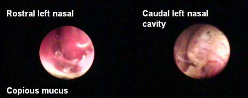
to clear mucoid discharge was present throughout the nasal cavity. Turbinates
in the rostral half of both nasal cavities appeared normal. In the caudal half
of the nasal cavities the surface of the mucosa was rough, irregular and friable.
In several areas blood vessels were prominent. No obvious fungal colonies or
masses were seen and no distinct localized area of abnormality was found.
Notes: A recent
CT scan of the skull indicated mild fluid accumulation bilaterally, right sphenoidal
sinusitis, swelling in the ventral meatus, periapcial disease of right 3rd and
4th upper premolars without extension into the nasal cavity and mild turbinate
destruction in the rostral right nasal cavity. The changes were bilateral and
reported as more severe in the right nasal cavity. This is in contrast to the
observed rhinoscopic findings wherein the changes were seen as more severe in
the caudal left nasal cavity. The final radiographic diagnosis was chronic rhinitis/sinusitis,
bilateral.
Hopefully,
the biopsy, culture samples and serologies will be revealing and lead to a final
diagnosis. Although neoplasia cannot be ruled out entirely, based on the gross
rhinoscopic findings fungal, allergic or idopathic rhinitis seem most likely.
|

